What Are the Different Compound Microscope Parts?
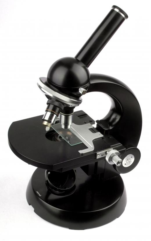
A compound microscope is an apparatus that uses a series of lenses to magnify the minute detail of a specimen that would otherwise not be visible with the naked eye. The compound microscope parts that are directly responsible for magnification include the objectives, the projector lens, and the ocular lenses or eye pieces. Illumination and concentration of the beam of light onto the specimen are provided by the light source, the iris diaphragm, or aperture and the condenser. The compound microscope parts responsible for focusing the image include the coarse and fine adjustment knobs. Supportive structures of the compound microscope include the slide stage, arm, and base.
Magnification of the specimen is the singular goal of a microscope, and the compound microscope parts that perform this task are the objectives, projection lens, and the ocular lenses. This enlargement is accomplished by concentrating a bright source of light onto a specimen from below. The light passes through the specimen and then into one of the objectives, where the lens magnifies the image. Most compound microscopes have two to four different objectives that are capable of magnifying the specimen to different degrees.

Passing light through the objective reverses the image. If the deviated light paths are directed next through the projector lens, the image is flipped back. A projector lens, however, is not necessary and an image can be viewed reversed without any loss of image quality.
Next, the light passes through the ocular lens, which typically magnifies the image another ten times and then projects the image into the user’s eye. Some microscopes have only one ocular lens while other microscopes have two. When a microscope has only one ocular lens, the magnified image is viewed with one eye and the other eye is closed.

The light source, iris diaphragm, and condenser are the compound microscope parts responsible for illuminating the specimen. The light source is simply a light bulb. Light from the bulb is passed through the aperture, a tool that can open and close to create a beam of light of different diameters. The beam of light then passes through the condenser, a lens that gathers and concentrates the light onto the specimen to maximize illumination.

Focus control of the image is provided by the compound microscope parts, called the course and fine adjustment knobs. These dials raise and lower the stage to bring the image into focus. The course adjustment knob moves the stage at a greater rate and is used first to bring the specimen close to focus. Final focus is done using the fine adjustment knob, which allows for tiny changes in the stage height and delicate control over the focus.
There are several parts of the compound microscope that are involved in structural support. The specimen is placed on the stage of the microscope and is held in place using clips. A U-shaped base provides a stable foundation for the entire microscope. The backbone of the microscope, supporting all the vertical components, is called the arm. A microscope is always carried by holding the arm and placing a hand under the base.
AS FEATURED ON:
AS FEATURED ON:


Discuss this Article
Post your comments