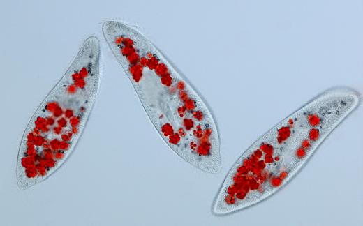
What is a Phase Contrast Microscope?
A phase contrast microscope is a scientific instrument specifically designed to increase the contrast of live specimens under observation. The microscope depends on the different refractive qualities of objects to distinguish between transparent and colorless structures. Other microscopy methods depend on staining a specimen to highlight or define different cellular components. The process of staining usually kills the specimen, making the study of active cellular processes impossible. The phase contrast microscope eliminates the need to kill a specimen by harnessing the nature of light waves.
A light wave contains peaks and valleys at regular intervals. If the peaks and valleys of different waves line up, they are said to be in phase. When they are misaligned the waves are out of phase.

The phase contrast microscope uses two light sources: a lamp under the specimen, and a light that is either diffracted or reflected off the specimen. Light is passed through a transparent object, while it is reflected off a solid, but colorless object. When the light waves are brought together in the phase condenser, a lens above the specimen, they will either be in phase or out of phase. If the light waves are in phase, the object will appear bright. If they are out of phase, the object will be shaded or dark.
Phase contrast microscopy was first developed around 1930 by Fritz Zerinke. His invention was not initially well received. When the German war machine caught hold of it in 1941, it was finally manufactured.

After the war, the phase contrast microscope continued to be made and applied to new areas of study, such as medicine. The phase contrast microscope has been instrumental in outlining the processes involved in cell division and other active cellular processes. Zerinke was later awarded the Nobel Prize in Physics in 1953 for his contribution to microscopy techniques.
AS FEATURED ON:
AS FEATURED ON:


Discuss this Article
Post your comments