What is Chromosome Banding?
Chromosome banding is the transverse bands that appear on chromosomes as a result of various differential staining techniques. Differential stains impart colors to tissues, so that they may be studied under a microscope. Chromosomes are thread-like structures of long deoxyribonucleic acid (DNA) filaments, which coil into a double helix and are made up of genetic information, or genes, that are arranged in a crosswise manner down the length.
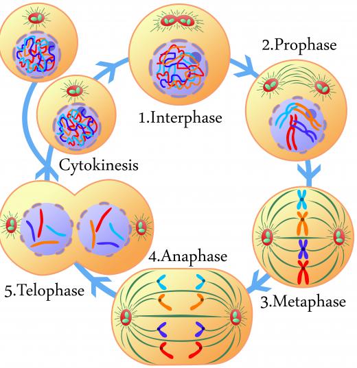
To analyze chromosomes under a microscope, they need to be stained when they are undergoing cell division during the meiosis or mitosis. Mitosis and meiosis are cell division processes that are divided into four phases. Those phases are prophase, metaphase, anaphase, and telophase.

Crytogenetics is the study of the function of cells, the structure of cells, DNA, and chromosomes. It employs various techniques for staining chromosomes, like G-banding, R-banding, C-banding, Q-banding, and T-banding. Each staining technique allows scientists to study different aspects of chromosome banding patterns.
Giemsa banding, also known as G-banding, enables scientists to study chromosomes in the metaphase stage of mitosis. Metaphase is the second stage of mitosis. At this phase the chromosomes are lined up and attached at the centers or their centromeres, and each chromosome appears in an X shape form.

Before applying stain to the chromosomes, they must first be treated with trypsin, which is a digestive fluid found in many animals. The trypsin will start to digest the chromosomes, allowing them to better receive the Giemsa stain. Giemsa stain was discovered by Gustav Giemsa, and is a mixture of methylene blue and the red acidic dye, eosin. Q-banding uses quinicrine, which is a mustard type solution. It produces results that are very similar to Giemsa, but has fluorescent qualities.

DNA is made up of four base acids that appear in pairs — adenine paired with thymine, and cytosine with guanine. Giemsa stain creates chromosome banding patterns with dark areas rich in adenine and thymine. The light areas are rich with guanine and cytosine. These areas replicate early and are euchromatic. Euchromatic is a genetically active area that stains very lightly with dye treatments.
Reverse-banding, or R-banding, produces chromosome banding patterns that are the opposite of G-banding. The darker areas are rich with guanine and cytosine. It also prodcues euchromatic parts with high concentrations of adenine and thymine.

With C-banding, the Giemsa stain is used to study the constitutive heterochromatin and the centromere of a chromosome. Constitutive heterochromatins are areas near the center of the chromosome that contain highly condensed DNA that tend to be transcriptionally silent. The centromere is the region at the very center of the chromosome.
T-banding allows scientists to study the telomeres of a chromosome. The telomeres are the caps that are on the each of the chromosomes. They contain repetitive DNA and are meant to prevent any deterioration from occurring.

Once the chromosomes are stained with Giemsa, researchers can clearly see the alternating dark and light chromosome banding patterns that are produced. By counting the number of bands, the karyotype of a cell can be determined. The karyotype is the characterization of chromosomes for a species according to size, type, and number.
AS FEATURED ON:
AS FEATURED ON:


Discuss this Article
Post your comments